Mind-controlled prosthetic arm moves individual ‘fingers’
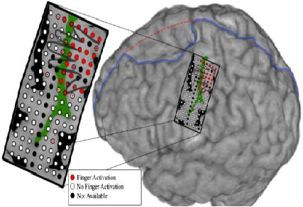
Posted by Acubiz | BlogAn illustration showing the electrode array on the subject’s brain, including a representation of what part of the brain controls each finger.
Physicians and biomedical engineers from Johns Hopkins report what they believe is the first successful effort to wiggle fingers individually and independently of each other using a mind-controlled artificial “arm” to control the movement.
The proof-of-concept feat, described online this week in the Journal of Neural Engineering, represents a potential advance in technologies to restore refined hand function to those who have lost arms to injury or disease, the researchers say. The young man on whom the experiment was performed was not missing an arm or hand, but he was outfitted with a device that essentially took advantage of a brain-mapping procedure to bypass control of his own arm and hand.
“We believe this is the first time a person using a mind-controlled prosthesis has immediately performed individual digit movements without extensive training,” says senior author Nathan Crone, M.D., professor of neurology at the Johns Hopkins University School of Medicine. “This technology goes beyond available prostheses, in which the artificial digits, or fingers, moved as a single unit to make a grabbing motion, like one used to grip a tennis ball.”
For the experiment, the research team recruited a young man with epilepsy already scheduled to undergo brain mapping at The Johns Hopkins Hospital’s Epilepsy Monitoring Unit to pinpoint the origin of his seizures.
While brain recordings were made using electrodes surgically implanted for clinical reasons, the signals also control a modular prosthetic limb developed by the Johns Hopkins University Applied Physics Laboratory.
Prior to connecting the prosthesis, the researchers mapped and tracked the specific parts of the subject’s brain responsible for moving each finger, then programmed the prosthesis to move the corresponding finger.
First, the patient’s neurosurgeon placed an array of 128 electrode sensors — all on a single rectangular sheet of film the size of a credit card — on the part of the man’s brain that normally controls hand and arm movements. Each sensor measured a circle of brain tissue 1 millimeter in diameter.
The computer program the Johns Hopkins team developed had the man move individual fingers on command and recorded which parts of the brain the “lit up” when each sensor detected an electric signal.
In addition to collecting data on the parts of brain involved in motor movement, the researchers measured electrical brain activity involved in tactile sensation. To do this, the subject was outfitted with a glove with small, vibrating buzzers in the fingertips, which went off individually in each finger. The researchers measured the resulting electrical activity in the brain for each finger connection.
After the motor and sensory data were collected, the researchers programmed the prosthetic arm to move corresponding fingers based on which part of the brain was active. The researchers turned on the prosthetic arm, which was wired to the patient through the brain electrodes, and asked the subject to “think” about individually moving thumb, index, middle, ring and pinkie fingers. The electrical activity generated in the brain moved the fingers.
“The electrodes used to measure brain activity in this study gave us better resolution of a large region of cortex than anything we’ve used before and allowed for more precise spatial mapping in the brain,” says Guy Hotson, graduate student and lead author of the study. “This precision is what allowed us to separate the control of individual fingers.”
Initially, the mind-controlled limb had an accuracy of 76 percent. Once the researchers coupled the ring and pinkie fingers together, the accuracy increased to 88 percent.
“The part of the brain that controls the pinkie and ring fingers overlaps, and most people move the two fingers together,” says Crone. “It makes sense that coupling these two fingers improved the accuracy.”
The researchers note there was no pre-training required for the subject to gain this level of control, and the entire experiment took less than two hours.
Crone cautions that application of this technology to those actually missing limbs is still some years off and will be costly, requiring extensive mapping and computer programming. According to the Amputee Coalition, over 100,000 people living in the U.S. have amputated hands or arms, and most could potentially benefit from such technology.
Scientists prove feasibility of ‘printing’ replacement tissue
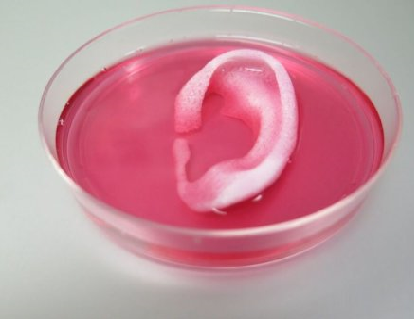
Posted by Acubiz | BlogCompleted ear structure printed with the Integrated Tissue-Organ Printing System.
Credit: Image courtesy of Wake Forest Baptist Medical Center
Using a sophisticated, custom-designed 3D printer, regenerative medicine scientists at Wake Forest Baptist Medical Center have proved that it is feasible to print living tissue structures to replace injured or diseased tissue in patients.
Reporting in Nature Biotechnology, the scientists said they printed ear, bone and muscle structures. When implanted in animals, the structures matured into functional tissue and developed a system of blood vessels. Most importantly, these early results indicate that the structures have the right size, strength and function for use in humans.
“This novel tissue and organ printer is an important advance in our quest to make replacement tissue for patients,” said Anthony Atala, M.D., director of the Wake Forest Institute for Regenerative Medicine (WFIRM) and senior author on the study. “It can fabricate stable, human-scale tissue of any shape. With further development, this technology could potentially be used to print living tissue and organ structures for surgical implantation.”
With funding from the Armed Forces Institute of Regenerative Medicine, a federally funded effort to apply regenerative medicine to battlefield injuries, Atala’s team aims to implant bioprinted muscle, cartilage and bone in patients in the future.
Tissue engineering is a science that aims to grow replacement tissues and organs in the laboratory to help solve the shortage of donated tissue available for transplants. The precision of 3D printing makes it a promising method for replicating the body’s complex tissues and organs. However, current printers based on jetting, extrusion and laser-induced forward transfer cannot produce structures with sufficient size or strength to implant in the body.
The Integrated Tissue and Organ Printing System (ITOP), developed over a 10-year period by scientists at the Institute for Regenerative Medicine, overcomes these challenges. The system deposits both bio-degradable, plastic-like materials to form the tissue “shape” and water-based gels that contain the cells. In addition, a strong, temporary outer structure is formed. The printing process does not harm the cells.
A major challenge of tissue engineering is ensuring that implanted structures live long enough to integrate with the body. The Wake Forest Baptist scientists addressed this in two ways. They optimized the water-based “ink” that holds the cells so that it promotes cell health and growth and they printed a lattice of micro-channels throughout the structures. These channels allow nutrients and oxygen from the body to diffuse into the structures and keep them live while they develop a system of blood vessels.
It has been previously shown that tissue structures without ready-made blood vessels must be smaller than 200 microns (0.007 inches) for cells to survive. In these studies, a baby-sized ear structure (1.5 inches) survived and showed signs of vascularization at one and two months after implantation.
“Our results indicate that the bio-ink combination we used, combined with the micro-channels, provides the right environment to keep the cells alive and to support cell and tissue growth,” said Atala.
Another advantage of the ITOP system is its ability to use data from CT and MRI scans to “tailor-make” tissue for patients. For a patient missing an ear, for example, the system could print a matching structure.
Several proof-of-concept experiments demonstrated the capabilities of ITOP. To show that ITOP can generate complex 3D structures, printed, human-sized external ears were implanted under the skin of mice. Two months later, the shape of the implanted ear was well-maintained and cartilage tissue and blood vessels had formed.
To demonstrate the ITOP can generate organized soft tissue structures, printed muscle tissue was implanted in rats. After two weeks, tests confirmed that the muscle was robust enough to maintain its structural characteristics, become vascularized and induce nerve formation.
And, to show that construction of a human-sized bone structure, jaw bone fragments were printed using human stem cells. The fragments were the size and shape needed for facial reconstruction in humans. To study the maturation of bioprinted bone in the body, printed segments of skull bone were implanted in rats. After five months, the bioprinted structures had formed vascularized bone tissue.
Ongoing studies will measure longer-term outcomes.
Parental Depression Negatively Affects Children’s School Performance
Posted by Acubiz | BlogA new study has found that when parents are diagnosed with depression, it can have a significant negative impact on their children’s performance at school.
Researchers at Drexel University led a team including faculty from the Karolinska Institutet in Stockholm, Sweden, and the University of Bristol in England in a cohort study of more than a million children born from 1984 until 1994 in Sweden. Using computerized data registers, the scientists linked parents’ depression diagnoses with their children’s final grades at age 16, when compulsory schooling ends in Sweden.
The research indicated that children whose mothers had been diagnosed with depression are likely to achieve grades that are 4.5 percentage points lower than peers whose mothers had not been diagnosed with depression. For children whose fathers were diagnosed with depression, the difference is a negative four percentage points.
Put into other terms, when compared with a student who achieved a 90 percent, a student whose mother or father had been diagnosed with depression would be more likely to achieve a score in the 85–86 percent range.
The magnitude of this effect was similar to the difference in school performance between children in low versus high-income families, but was smaller than the difference for low versus high maternal education (low family income: -3.6 percentage points; low maternal education -16.2 percentage points).
How well a student does in school has a large bearing on future job and income opportunities, which has heavy public health implications, explained Félice Lê-Scherban, PhD, assistant professor in the Dornsife School of Public Health. On average in the United States, she said, an adult without a high school degree earns half as much as one of their peers with a college degree and also has a life expectancy that is about 10 years lower.
“Anything that creates an uneven playing field for children in terms of their education can potentially have strong implications for health inequities down the road,” Lê-Scherban said.
Some differences along gender lines were observed in the study. Although results were largely similar for maternal and paternal depression, analysis found that episodes of depression in mothers when their children were 11–16 years old appeared to have a larger effect on girls than boys. Girls scored 5.1 percentage points lower than their peers on final grades at 16 years old when that factor was taken into account. Boys, meanwhile, only scored 3.4 percentage points lower.
Brian Lee, PhD, associate professor in the Dornsife School of Public Health, said there were gender differences in the study’s numbers, but didn’t want to lose focus of the problem parental depression presents as a whole.
“Our study — as well as many others — supports that both maternal and paternal depression may independently and negatively influence child development,” Lee said. “There are many notable sex differences in depression, but, rather than comparing maternal versus paternal depression, we should recognize that parental depression can have adverse consequences not just for the parents but also for their children.”
Depression diagnoses in a parent at any time during the child’s first 16 years were determined to have some effect on the child’s school performance. Even diagnoses of depression that came before the child’s birth were linked to poorer school performance. The study posited that it could be attributed to parents and children sharing the same genes and the possibility of passing on a disposition for depression.
The study, “Associations of Parental Depression With Child School Performance at Age 16 Years in Sweden,” whose lead author was Drexel alumna Hanyang Shen, was published in JAMA Psychiatry
NIH scientists discover genetic cause of rare allergy to vibration
Posted by Acubiz | BlogScientists at the National Institutes of Health (NIH) have identified a genetic mutation responsible for a rare form of inherited hives induced by vibration, also known as vibratory urticaria. Running, hand clapping, towel drying or even taking a bumpy bus ride can cause temporary skin rashes in people with this rare disorder. By studying affected families, researchers discovered how vibration promotes the release of inflammatory chemicals from the immune system’s mast cells, causing hives and other allergic symptoms.
Their findings, published online in the New England Journal of Medicine on Feb. 3, suggest that people with this form of vibratory urticaria experience an exaggerated version of a normal cellular response to vibration. The study was led by researchers at the National Institute of Allergy and Infectious Diseases (NIAID) and the National Human Genome Research Institute (NHGRI), both part of NIH.
“Investigating rare disorders such as vibratory urticaria can yield important insights into how the immune system functions and how it reacts to certain triggers to produce allergy symptoms, which can range from mild to debilitating,” said NIAID Director Anthony S. Fauci, M.D. “The findings from this study uncover intriguing new facets of mast cell biology, adding to our knowledge of how allergic responses occur.”
“This study illustrates the power of a multidisciplinary team, involving clinicians, geneticists and basic immunologists, to get to the heart of a medical mystery,” said Dan Kastner, M.D., Ph.D., scientific director of the Intramural Research Program at NHGRI and a co-author of the study. “It also underscores the tremendous potential of new genomic techniques.”
In addition to itchy red welts at the site of vibration on the skin, people with vibratory urticaria also sometimes experience flushing, headaches, fatigue, blurry vision or a metallic taste in the mouth. Symptoms usually disappear within an hour, but those affected may experience several episodes per day.
The current study involved three families in which multiple generations experienced vibratory urticaria. The NIH team evaluated the first family under an ongoing clinical protocol investigating urticarias induced by a physical trigger.
Mast cells, which reside in the skin and other tissues, release histamine and other inflammatory chemicals into the bloodstream and surrounding tissue in response to certain stimuli, a process known as degranulation. To assess potential mast cell involvement in vibratory urticaria, the researchers measured blood levels of histamine during an episode of vibration-induced hives. Histamine levels rose rapidly in response to vibration and subsided after about an hour, indicating that mast cells had released their contents. The researchers also observed increased tryptase, another marker of mast cell degranulation, in skin around the affected area.
“Notably, we also observed a small increase in blood histamine levels and a slight release of tryptase from mast cells in the skin of unaffected individuals exposed to vibration,” said Hirsh Komarow, M.D., of NIAID’s Laboratory of Allergic Diseases, the senior author of the study. “This suggests that a normal response to vibration, which does not cause symptoms in most people, is exaggerated in our patients with this inherited form of vibratory urticaria.”
The NIH team realized that the first family’s symptoms matched those of a different family described by researchers at Yale University in 1981. Through a collaboration with Yale, the NIH team obtained DNA samples from 25 members of that family. Two family members came to NIH for evaluation, and they put the scientists in contact with a third family with similar symptoms.
To identify the genetic basis of the disorder, the scientists performed genetic analyses, including DNA sequencing, on 36 affected and unaffected members from the three families. They found a single mutation in the ADGRE2 gene shared by family members with vibratory urticaria but not present in unaffected people. The scientists did not detect the ADGRE2 mutation in variant databases or in the DNA of more than 1,000 unaffected individuals with a similar genetic ancestry as the three families.
“This work marks, to the best of our knowledge, the first identification of a genetic basis for a mast-cell-mediated urticaria induced by a mechanical stimulus,” said Dean Metcalfe, M.D., chief of NIAID’s Laboratory of Allergic Diseases and a study co-author.
The ADGRE2 gene provides instructions for production of ADGRE2 protein, which is present on the surface of several types of immune cells, including mast cells. ADGRE2 is composed of two subunits–a beta subunit located within the cell’s outer membrane, and an alpha subunit located on the outside surface of the cell. Normally, these two subunits interact, staying close together.
People with familial vibratory urticaria produce a mutated ADGRE2 protein in which this subunit interaction is less stable, the investigators found. After vibration, the alpha subunit of the mutant protein was no longer in close contact with the beta subunit. When the alpha subunit detaches from the beta subunit, the researchers suggest, the beta subunit produces signals inside mast cells that lead to degranulation, which causes hives and other allergy symptoms.
The research suggests that the ADGRE2 subunit interaction plays a key role in the mast cell response to certain physical stimuli, which could have implications for other diseases in which mast cells are involved. Next, the scientists plan to study what happens to the alpha subunit post-vibration and to unravel the cellular signaling leading to degranulation. They also plan to recruit more families with vibratory urticaria to further study the disorder and look for additional mutations in ADGRE2 and other genes.
Breakthrough in generating embryonic cells that are critical for human health
Posted by Acubiz | BlogNeural crest cells arise early in the development of vertebrates, migrate extensively through the embryo, and differentiate to give rise to a wide array of diverse derivatives. Their contributions include a large proportion of our peripheral nerves, the melanocytes that provide skin color and protection from damaging UV light, as well as many different cell types in our face, including muscle, bone, cartilage and tooth-forming cells.
The proper functioning of these cells is critical for human development and health. When neural crest biology fails, various birth defects and illnesses – cleft lip/palate, Hirschsprung and Waardenburgsyndromes, melanoma and neuroblastoma – result. A better study of these cells is crucial, therefore, to aid in clinical efforts to diagnose and treat such conditions.
But access to these embryonic cells in humans is very difficult. As an alternative, scientists turned to models based in embryonic stem cells.
While protocols to generate human neural crest cells from human embryonic stem cells have progressed since the first report 11 years ago, they still have considerable limitations for their use in basic and clinical research. This is because these protocols commonly use ingredients or components not well defined, such as blood serum which contains many unknown components of varying concentrations. Some protocols result in large clusters of cells, impairing the identification of specific molecules and their roles during neural crest cell formation. Furthermore, the fastest of these protocols takes 12 days (of very costly culture conditions) to convert human embryonic stem cells to neural crest cells. Oftentimes the protocols provide low yields, making the isolation of the desired neural crest cells a time-consuming and technically challenging process.
Work done by a research team led by an associate professor of biomedical sciences in the School of Medicine at the University of California, Riverside now addresses these problems by providing a robust, fast, simple and trackable method to generate neural crest cells. The proposed method can facilitate research in basic sciences and clinical applications alike.
“Our study provides a superb model to generate neural crest cells in just five days starting from human embryonic stem cells or induced pluripotent cells, using a simple and well-defined media with all ingredients known and accounted for,” said Martín I. García-Castro, whose lab led the studypublished in the Feb. 1 issue of the journal Development. “Our cost-effective, efficient and fast protocol allows a better analysis of the relevant signals and molecules involved in the formation of these cells. Our results suggest that human neural crest cells can arise independently from – and prior to – the formation of mesoderm and neural ectoderm derivatives, both of which had been thought to be critical for neural crest formation.”
The mesoderm is the middle layer of the embryo in early development. It lies between the endoderm and the ectoderm, with the latter being the outermost layer. García-Castro’s previous work on birds already challenged the dogma suggesting that neural crest cells form without mesodermal or neural contribution. Unpublished results from his lab have also confirmed the same using rabbit embryos as a mammalian model.
With regard to identifying specific molecules and their roles during neural crest cell formation, García-Castro’s new work demonstrates the critical role played by a molecule known as WNT and highlights contributions from protein families called FGFs and BMPs.
Briefly, WNT proteins are signaling molecules that regulate cell-to-cell interactions during development and adult tissue homeostasis. The FGF protein family controls a wide range of biological functions. BMPs induce the formation of bone and cartilage and form tissues throughout the body.
“Our work provides strong evidence of the critical and initiating role of WNT signals in neural crest cell formation, with later contributions by FGF and BMP pathways,” he said.
García-Castro emphasized that the proper function of neural crest cells is essential for human development and health.
“The study of these cells is essential to improve clinical efforts to diagnose, manage, and perhaps prevent diseases and conditions linked to them, and our lab has already launched efforts towards facial clefts – lip and or palate – and melanoma, and we hope to make substantial progress in both areas thanks to this novel protocol,” he said.
Your Facial Bone Structure Has a Big Influence on How People See You
Posted by Acubiz | BlogNew research shows that although we perceive character traits like trustworthiness based on a person’s facial expressions, our perceptions of abilities like strength are influenced by facial structure
By Jessica Schmerler on June 18, 2015
Selfies, headshots, mug shots — photos of oneself convey more these days than snapshots ever did back in the Kodak era. Most digitally minded people continually post and update pictures of themselves at professional, social media and dating sites such as LinkedIn, Facebook, Match.com and Tinder. For better or worse, viewers then tend to make snap judgments about someone’s personality or character from a single shot. As such, it can be a stressful task to select the photo that conveys the best impression of ourselves. For those of us seeking to appear friendly and trustworthy to others, a new study underscores an old, chipper piece of advice: Put on a happy face.
A newly published series of experiments by cognitive neuroscientists at New York University is reinforcing the relevance of facial expressions to perceptions of characteristics such as trustworthiness and friendliness. More importantly, the research also revealed the unexpected finding that perceptions of abilities such as physical strength are not dependent on facial expressions but rather on facial bone structure.
The team’s first experiment featured photographs of 10 different people presenting five different facial expressions each. Study subjects rated how friendly, trustworthy or strong the person in each photo appeared. A separate group of subjects scored each face on an emotional scale from “very angry” to “very happy.” And three experts not involved in either of the previous two ratings to avoid confounding results calculated the facial width-to-height ratio for each face. An analysis revealed that participants generally ranked people with a happy expression as friendly and trustworthy but not those with angry expressions. Surprisingly, participants did not rank faces as indicative of physical strength based on facial expression but graded faces that were very broad as that of a strong individual.
In a second survey facial expression and facial structure were manipulated in computer-generated faces. Participants rated each face for the same traits as in the first survey, with the addition of a rating for warmth. Again, people thought a happy expression, but not an angry one, indicated friendliness, trustworthiness — and in this case, warmth. The researchers then showed two additional sets of participants the same faces, this time either with areas relevant to facial expressions obscured or the width cropped. In the first variation, for faces lacking emotional cues, people could no longer perceive personality traits but could still perceive strength based on width. Similarly, for those faces lacking structural cues, people could no longer perceive strength but could still perceive personality traits based on facial expressions.
In a third iteration of the survey participants had to pick four faces out of a lineup of eight faces varied for expression and width that they might select either as their financial advisor or as the winner of a power-lifting competition. As might be expected, participants picked faces with happier expressions as financial advisors and selected broader faces as belonging to power-lifting champs.
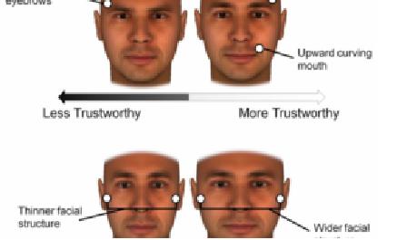
In a final survey the researchers generated more than 100 variations of one individual “base face” by varying facial features. Participants saw two faces at a time, and then picked one as either trustworthy or high in ability or as a good financial advisor or power-lifting winner. Using these results, a computer then created an average face for each of these four categories, which were shown to a separate set of participants who had to pick which face appeared either more trustworthy or stronger. Most of the participants found the computer-generated averages to be good representations of trustworthiness or strength — and generally saw the average “financial advisor” face as more trustworthy and the “power-lifter” face as stronger. The findings from all four surveys were published in the Personality and Social Psychology Bulletin on June 18.
Taken together the findings suggest facial expressions strongly influence perception of traits such as trustworthiness, friendliness or warmth, but not ability (strength, in these experiments). Conversely, facial structure influences the perception of physical ability but not intentions (such as friendliness and trustworthiness, in this instance). In addition, decisions that involve guessing at the possible intentions of a person such as to whom you would entrust your money management are more strongly influenced by facial expression, whereas those based on physical ability such as whom you would bet on in a sporting event are more strongly influenced by facial structure.
Previous studies also have shown the effect of facial cues on how we perceive and interact with others but this new work reveals how perceptions of the same person can vary greatly depending on that person’s facial expression in any given moment. This variability “has implications for both the people presenting themselves and the perceivers in social interactions,” says Jonathan Freeman, a social neuroscientist at New York University and senior author of the study. So, we might consider the impact of our facial expressions in the photos we post online. At the same time, in an ideal world people who look at our photos would give us the benefit of the doubt and hesitate to make spontaneous judgments based only on a single image.
The findings above come with a big caveat: Only male faces were shown to subjects. The researchers chose this approach because previous studies involving the ratio of facial width to height have shown that greater facial width is often associated with higher testosterone levels as well asheightened aggression and strength in men. Studies of facial width and height in females have shown mixed results, so presenting study subjects with a mix of male and female faces would have yielded inconclusive results. Despite the relative lack of evidence on how facial structure influences perception of women’s faces, there have been humorous portrayals of popular speculations. Future research, however, is needed to definitively establish whether any such patterns exist.
Furthermore, the researchers refer to “ability” when discussing physical strength in the study. No specific measurements were made, for example, of perceptions of intellectual ability or ability to perform in certain job positions. These abilities are more abstract and thus might rely on a combination of different dynamic and static facial cues, Freeman explains, so it would be difficult to test these relationships definitively.
In our everyday lives this study and others make clear that although we might try to influence others’ perceptions of us with photos showing us donning sharp attire or displaying a self-assured attitude, the most important determinant of others’ perception of and consequent behaviortoward us is our faces.
So the next time you want to win someone’s trust, try a smile and a happy face. But for those folks hoping to get picked for a pick-up game of football, basketball and so on, don’t worry about your facial expression. The best you can do is hope you have a wider face and then let your physical prowess speak for itself.
Gene therapy pioneer James Wilson uses CRISPR/Cas9 to target liver disease
Posted by Acubiz | BlogOne of the pioneers in the whole gene therapy movement of the past 35 years has combined his knowledge of viral vectors with the hot new CRISPR/Cas9 tech to tackle a rare genetic liver disease. And his work with rodents highlighted both the promise of this new technology as well as an unexpected hurdle.
James Wilson at the University of Pennsylvania is virtually the father of adeno-associated virus–or AAV–vectors used to deliver gene therapies. After years of up and down progress, including an early patient death that slowed research in gene therapies for decades, his technology was in-licensed by ReGenX, which has since outlicensed it to a group of biotechs pursuing new gene therapy programs.
In what is described as a first in research circles, he used an AAV vector to deliver the components of CRISPR/Cas9 gene editing tools into a mouse model of a rare metabolic urea-cycle disorder, triggered by a deficiency in the ornithine transcarbamylase (OTC) enzyme. When one of a series of enzymes is missing or “deficient,” ammonia can accumulate in the blood and circulate into the brain, where it can badly damage brains. OTC deficiency occurs in one in every 40,000 births.
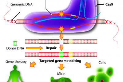
Wilson’s group used one AAV to deliver a Cas9 enzyme–the cutting tool–specifically into liver cells. Another vector carried a guide RNA to the specified spot and donor DNA to correct the mutation in a cut-and-paste approach that is spreading around academic labs like wild fire.
In a research article published in Nature Biotechnology, Wilson says his team achieved a 10% reversion in the liver cells of newborn mice, with an increased survival rate for challenged rodents and a high death rate in the untreated group of newly born mice. In adult mice, though, the team say that the experiment didn’t work as planned, highlighting a needed correction in the process. And now they’re following up with new approaches to see how that might work.
“Correcting a disease-causing mutation following birth in this animal model brings us one step closer to realizing the potential of personalized medicine,” said Wilson, a professor of medicine and director of the Orphan Disease Center at Penn. “Nevertheless, my 35-year career in gene therapy has taught me how difficult translating mouse studies to successful human treatments can be. From this study, we are now adjusting the gene-editing system in the next phases of our investigation to address the unforeseen complications seen in adult animals.”
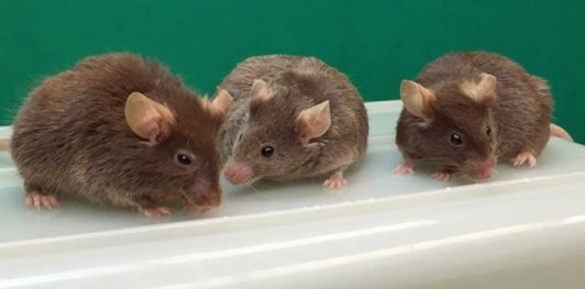
Male mice without any Y chromosome genes can father offspring after assisted reproduction
Posted by Acubiz | BlogThree males lacking any Y chromosome genes produced by ROSI. The males shown on the left and right are 2 years and 1 month old, and the male in the center is 1 year and 10 months old.
Credit: Yasuhiro Yamauchi
The Y chromosome is a symbol of maleness, present only in males and encoding genes important for male reproduction. But a new study has shown that live mouse progeny can be generated with assisted reproduction using germ cells from males which do not have any Y chromosome genes. This discovery adds a new light to discussions on Y chromosome gene function and evolution. It supports the hypothesis that Y chromosome genes can be replaced by that encoded on other chromosomes.
Two years ago, the University of Hawaii (UH) team led by Monika A. Ward, Professor at the Institute for Biogenesis Research, John A. Burns School of Medicine, University of Hawai’i, demonstrated that only two genes of the Y chromosome, the testis determinant factor Sry and the spermatogonial proliferation factor Eif2s3y, were needed for male mice to sire offspring with assisted fertilization. Now, the same team, with a collaborating researcher from France, Michael Mitchell (INSERM, Marseille), took a step further and produced males completely devoid of the entire Y chromosome.
In this new study scheduled for online publication in the journal Science on Jan. 29, 2016, Ward and her UH colleagues describe how they generated the “No Y” males, and define the ability of these males to produce gametes and sire offspring.
The UH researchers first replaced the Y chromosome gene Sry with its homologue and direct target encoded on chromosome 11, Sox9. In normal situation, Sry activates Sox9, and this initiates a cascade of molecular events that ultimately allow an XY fetus to develop into a male. The researchers used transgenic technology to activate Sox9 in the absence of Sry.
Next, they replaced the second essential Y chromosome gene, Eif2s3y, with its X chromosome encoded homologue, Eif2s3x. Eif2s3y and Eif2s3x belong to the same gene family and are very similar in sequence. The researchers speculated that these two genes may play similar roles, and it is a global dosage of both that matters. They transgenically overexpressed Eif2s3x, increasing dose of the X gene beyond that provided normally by X and Y. Under these conditions, Eif2s3x took over the function of Eif2s3y in initiating spermatogenesis.
Finally, Ward’s team replaced Sry and Eif2s3y simultaneously, and created XOSox9,Eif2s3x males that had no Y chromosome DNA. Mice lacking all Y chromosome genes developed testes populated with male germ cells. Round spermatids were harvested and a technique called round spermatid injection (ROSI) was used to successfully fertilize oocytes. When the developed embryos where transferred to female mouse surrogate mothers, live offspring were born.
The offspring derived from the “No Y” males were healthy and lived for normal life span. The daughters and grandsons of the “No Y” males were fertile and capable of reproducing on its own without further technological intervention. Ward’s team produced three consecutive generations of “No Y” males using ROSI showing that males lacking Y chromosome genes can be repeatedly propagated with technical assistance.
“Most of the mouse Y chromosome genes are necessary for development of mature sperm and normal fertilization, both in mice and in humans,” Ward said. “However, when it comes to assisted reproduction, we have now shown that in the mouse the Y chromosome contribution is not necessary.”
The study provides new important insights into Y chromosome gene function and evolution. It supports the existence of functional redundancy between the Y chromosome genes and their homologues encoded on other chromosomes. “This is good news,” Ward said, “because it suggests that there are back-up strategies within genomes, which are normally silent but are capable of taking over under certain circumstances. We revealed two of these strategies by genome manipulation. Whether such alternative pathways would ever be activated without human help, for example in response to environmental changes, is unknown. But it is certainly possible and has already happened for two rodent species which lost their Y chromosomes. ”
The development of assisted reproduction technologies (ART) allows bypassing various steps of normal fertilization by using immotile, non-viable, or immature gametes. The newest study as well as Ward’s preceding report (Science 2014 Jan 3; 343 (6166: 69-72) support that in the mouse ROSI is a successful and efficient form of ART. In humans, ROSI is considered experimental due to concerns regarding the safety of injecting immature germ cells and other technical difficulties. The researchers hope that the success in mouse studies may spark the re-evaluation of human ROSI for its suitability to become an option for overcoming male infertility in the future.
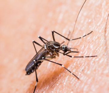
Zika Virus
Posted by Acubiz | BlogOn Monday (Jan. 25), the World Health Organization announced that Zika virus, a mosquito-borne illness that in the past year has swept quickly throughout equatorial countries, is expected to spread across the Americas and into the United States.
The disease, which was discovered in 1947 but had since been seen in only small, short-lived outbreaks, causes symptoms including a rash, headache and small fever. However, a May 2015 outbreak in Brazil led to nearly 3,500 reports of birth defects linked to the virus, even after its symptoms had passed, and an uptick in cases of Guillain-Barre syndrome, an immune disorder. The Centers for Disease Control and Prevention has issued a travel alert advising pregnant women to avoid traveling to countries where the disease has been recorded.
Zika virus is transmitted by the mosquito species Aedes aegypti, also a carrier of dengue fever and chikungunya, two other tropical diseases. Though Aedes aegypti is not native to North America, researchers at the University of Notre Dame who study the species have reported a discovery of a population of the mosquitoes in a Capitol Hill neighborhood in Washington, D.C. To add insult to injury, the team identified genetic evidence that these mosquitoes have overwintered for at least the past four years, meaning they are adapting for persistence in a northern climate well out of their normal range.
While the Washington population is currently disease-free, Notre Dame Department of Biological Sciences professor David Severson, who led the team, noted that the ability of this species to survive in a northern climate is troublesome. This mosquito is typically restricted to tropical and subtropical regions of the world and not found farther north in the United States than Alabama, Mississippi, Georgia and South Carolina.
“What this means for the scientific world,” said Severson, who led the team, “is some mosquito species are finding ways to survive in normally restrictive environments by taking advantage of underground refugia. Therefore, a real potential exists for active transmission of mosquito-borne tropical diseases in popular places like the National Mall. Hopefully, politicians will take notice of events like this in their own backyard and work to increase funding levels on mosquitoes and mosquito-borne diseases.”
Severson’s research focuses on mosquito genetics and genomics with a primary goal of understanding disease transmission. He has studied and tracked mosquitoes all over the world and most recently served as the director of the Eck Institute for Global Health at Notre Dame. His team, in coordination with the Disease Carrying Insects Program of Fairfax County Health Department in Fairfax, Virginia, recently published their findings in the American Journal of Tropical Medicine and Hygiene.
Notre Dame has a long history of mosquito research, studying both Aedes aegypti and Anopheles gambiae species, vector control and using mathematical models to better understand the dynamics of infectious disease transmission and control. Alex Perkins, Eck Family Assistant Professor of Biological Sciences, focuses on using mathematical, statistical and computational approaches to study mosquito-borne pathogens including dengue, chikungunya and Zika. Perkins uses the models to understand how to best control and prevent transmission of these diseases. He has previously worked with the CDC on making recommendations for chikungunya and dengue virus, and said he has discussed working with the CDC on Zika virus modeling.
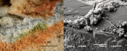
Antarctic fungi survive Martian conditions on the International Space Station
Posted by Acubiz | BlogDate: January 28, 2016
Source: FECYT – Spanish Foundation for Science and Technology
Summary:
Scientists have gathered tiny fungi that take shelter in Antarctic rocks and sent them to the International Space Station. After 18 months on board in conditions similar to those on Mars, more than 60 percent of their cells remained intact, with stable DNA. The results provide new information for the search for life on the red planet. Lichens from the Sierra de Gredos (Spain) and the Alps (Austria) also traveled into space for the same experiment.
Credit: S. Onofri et al.
European scientists have gathered tiny fungi that take shelter in Antarctic rocks and sent them to the International Space Station. After 18 months on board in conditions similar to those on Mars, more than 60% of their cells remained intact, with stable DNA. The results provide new information for the search for life on the red planet. Lichens from the Sierra de Gredos (Spain) and the Alps (Austria) also travelled into space for the same experiment.
The McMurdo Dry Valleys, located in the Antarctic Victoria Land, are considered to be the most similar earthly equivalent to Mars. They make up one of the driest and most hostile environments on our planet, where strong winds scour away even snow and ice. Only so-called cryptoendolithic microorganisms, capable of surviving in cracks in rocks, and certain lichens can withstand such harsh climatological conditions.
A few years ago a team of European researchers travelled to these remote valleys to collect samples of two species of cryptoendolithic fungi: Cryomyces antarcticus and Cryomyces minteri. The aim was to send them to the International Space Station (ISS) for them to be subjected to Martian conditions and space to observe their responses.
The tiny fungi were placed in cells (1.4 centimetres in diameter) on a platform for experiments known as EXPOSE-E, developed by the European Space Agency to withstand extreme environments. The platform was sent in the Space Shuttle Atlantis to the ISS and placed outside the Columbus module with the help of an astronaut from the team led by Belgian Frank de Winne.
For 18 months half of the Antarctic fungi were exposed to Mars-like conditions. More specifically, this is an atmosphere with 95% CO2, 1.6% argon, 0.15% oxygen, 2.7% nitrogen and 370 parts per million of H2O; and a pressure of 1,000 pascals. Through optical filters, samples were subjected to ultra-violet radiation as if on Mars (higher than 200 nanometres) and others to lower radiation, including separate control samples.
“The most relevant outcome was that more than 60% of the cells of the endolithic communities studied remained intact after ‘exposure to Mars’, or rather, the stability of their cellular DNA was still high,” highlights Rosa de la Torre Noetzel from Spain’s National Institute of Aerospace Technology (INTA), co-researcher on the project.
The scientist explains that this work, published in the journal Astrobiology, forms part of an experiment known as the Lichens and Fungi Experiment (LIFE), “with which we have studied the fate or destiny of various communities of lithic organisms during a long-term voyage into space on the EXPOSE-E platform.”
“The results help to assess the survival ability and long-term stability of microorganisms and bioindicators on the surface of Mars, information which becomes fundamental and relevant for future experiments centred around the search for life on the red planet,” states De la Torre.
Also lichens from Gredos and the Alps
Researchers from the LIFE experiment, coordinated from Italy by Professor Silvano Onofri from the University of Tuscany, have also studied two species of lichens (Rhizocarpon geographicum and Xanthoria elegans) which can withstand extreme high-mountain environments. These have been gathered from the Sierra de Gredos (Avila, Spain) and the Alps (Austria), with half of the specimens also being exposed to Martian conditions.
Another range of samples (both lichens and fungi) was subjected to an extreme space environment (with temperature fluctuations of between -21.5 and +59.6 ºC, galactic-cosmic radiation of up to 190 megagrays, and a vacuum of between 10-7 to 10-4 pascals). The effect of the impact of ultra-violet extraterrestrial radiation on half of the samples was also examined.
After the year-and-a-half-long voyage, and the beginning of the experiment on Earth, the two species of lichens ‘exposed to Mars’ showed double the metabolic activity of those that had been subjected to space conditions, even reaching 80% more in the case of the species Xanthoria elegans.
The results showed subdued photosynthetic activity or viability in the lichens exposed to the harsh conditions of space (2.5% of samples), similar to that presented by the fungal cells (4.11%). In this space environment, 35% of fungal cells were also seen to have kept their membranes intact, a further sign of the resistance of Antarctic fungi.